Skip to content

For Nigerian diagnostic centres, hospitals and HMOs

The ECG is printed. The heart doctor isn't here. TraceRead reads it first.

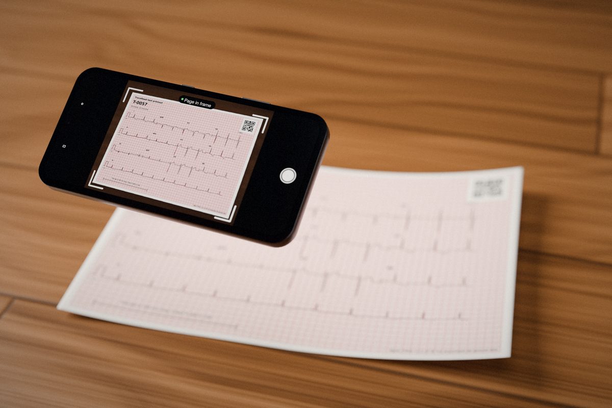
Take a photo of the paper ECG. Seconds after it arrives, your doctor has a first read to check: the heart rate, the key intervals and the patterns it sees. Your doctor still decides.

What TraceRead is, in plain words

01

The situation

Nigeria has about 400 cardiologists (heart doctors) for more than 220 million people, according to the Nigerian Cardiac Society. Many ECGs are read by doctors who are not heart specialists, or they wait for one.

02

What it costs you

Printouts sit in a pile until a heart doctor is free. Patients wait for a result or come back another day. The doctor on duty reads alone.

03

What TraceRead does

You photograph the printout. Seconds after the photo arrives, TraceRead AI has rebuilt the 12 tracings, measured them and listed the patterns it sees, each with a score from 0 to 100.

04

Who decides

Your doctor. TraceRead signs nothing, and nothing reaches a patient until a doctor has checked and signed it.

Heart doctor numbers: President of the Nigerian Cardiac Society, quoted in The Guardian (Nigeria).

A real first read, replayed

Watch a first read come together.

The numbers are TraceRead's own measures of three public research ECGs, printed and photographed by our simulator. They replay as the phone reads each page.

Every step of a real run
Heart rate63a min
PR interval136ms
QRS width88ms
QT interval406ms
QTc414ms
Heart axis20deg
Trace match0.97
Pulses read4 of 4
Page layout3x4
CheckPulse width

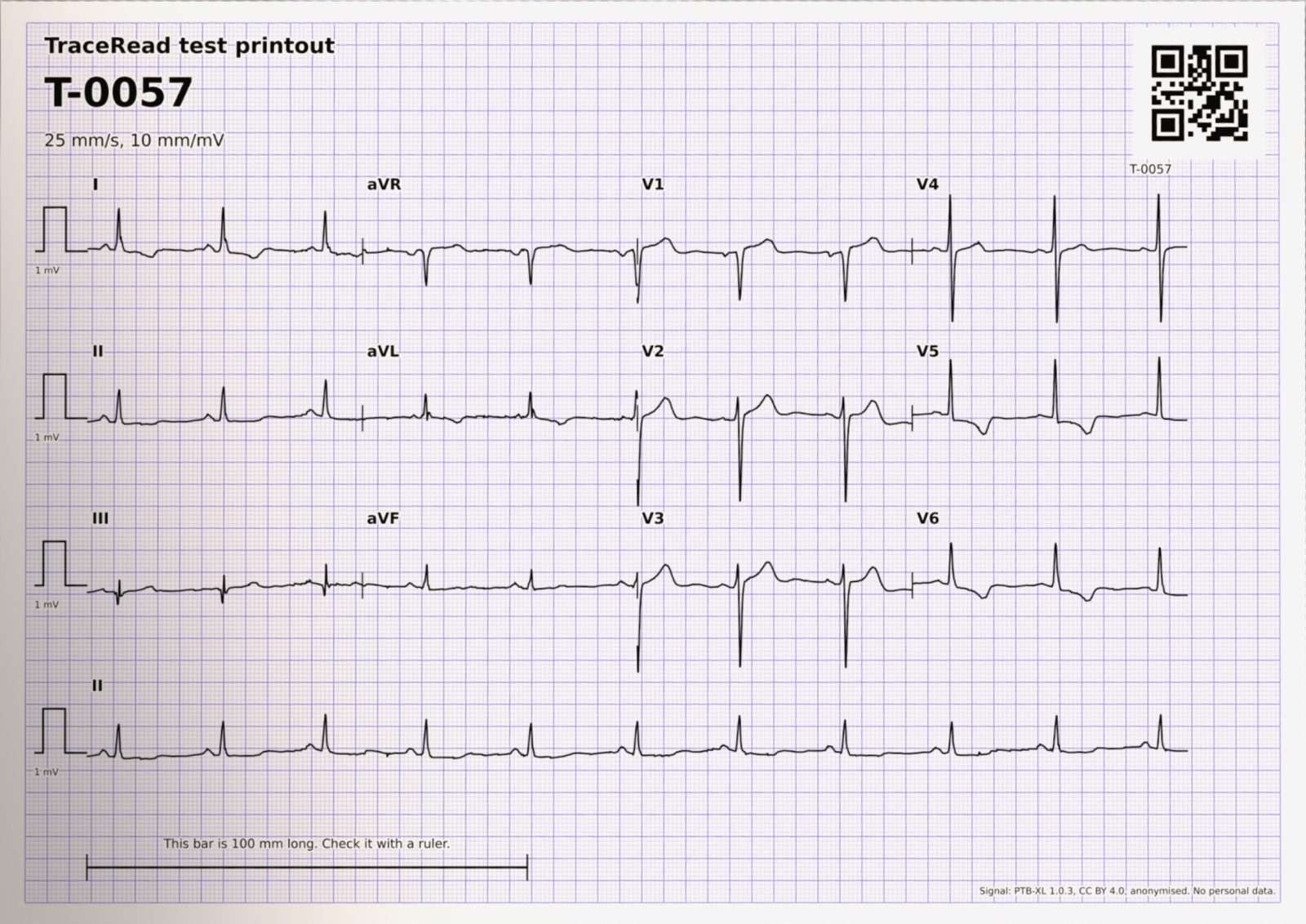
Run T-0057, from PTB-XL (Germany)

Lead II strip0.0 s

The AI first read

See exactly what your doctor gets back.

Real runs of TraceRead's code on public research ECGs from PTB-XL. Hover a lead to see how closely the red rebuilt line follows the original recording.

Flattened page of PTB-XL record 15863

Numbers under each lead: how closely the rebuilt line follows the original recording, where 1.00 is a perfect match. Median for this page: 0.96.

Photo usableAI first read, for a doctor to check

PTB-XL record 15863, a man aged 68. A4 paper, 3x4 with one lead II strip, pulses read 4 of 4.

Measures

MeasureFrom the photoOriginal recording
Heart rate (beats a minute)6363
PR interval (ms)136137
QRS width (ms)8887
QT interval (ms)406396
QTc, corrected for heart rate (ms)414404
Heart axis (degrees)2019

Patterns flagged, with TraceRead AI's score

  • Sinus rhythm

    the usual rhythm, started by the heart's natural pacemaker

    95
  • Left ventricular hypertrophy

    the muscle wall of the main pumping chamber may be thicker than usual

    60
  • Unspecific reduced blood flow pattern

    beat changes that can go with reduced blood flow to the heart muscle, without a clear place

    56
  • Reduced blood flow pattern, front and side

    beat changes over the front and side of the heart that can go with reduced blood flow to the heart muscle

    27
  • ST depression

    the middle part of the beat sits lower than usual

    19
  • Unspecific wide QRS

    the signal spreads slowly inside the lower chambers, so the main spike is wide

    7
  • Left atrial enlargement

    the upper left chamber may be larger or under more strain than usual

    2

What the dataset's cardiologists recorded for this ECG

  • Unspecific reduced blood flow pattern
  • Left ventricular hypertrophy
  • Sinus rhythm

Case studies

Twelve real ECGs, read end to end.

Page T-0057 with the rebuilt trace in red
T-0057, found every statement

Twelve ECGs, each read end to end

▲▼
0.908median match, rebuilt trace against the original recording500 photos
99.0%page layouts read right500 photos
0.902AUROC across 71 patterns2,198 ECGs
20 of 29patterns at 0.90 or more on ECGs from China45,152 ECGs
0.009AUROC lost reading photos instead of recordings500 photos
16.8 msaverage QTc difference from a reference program2,198 ECGs
7.85 sto read 95 in 100 ECGs on a 2 vCPU server97 photos
0.908median match, rebuilt trace against the original recording500 photos
99.0%page layouts read right500 photos
0.902AUROC across 71 patterns2,198 ECGs
20 of 29patterns at 0.90 or more on ECGs from China45,152 ECGs
0.009AUROC lost reading photos instead of recordings500 photos
16.8 msaverage QTc difference from a reference program2,198 ECGs
7.85 sto read 95 in 100 ECGs on a 2 vCPU server97 photos

What TraceRead does

Ten things TraceRead does between the photo and the first read.

For each one: what it does, what code checks without relying on the AI, and what a person does.

Stage 1

Capture

On the phone, before anything is sent

Photo check

The phone checks the picture about five times a second for blur, glare, poor light and a page that does not fill the frame, and says what to fix.

What code checks
In testing on 522 frames it flagged every one with blur of half a millimetre or more, large glare, poor light or a page too small, and none of 69 good ones.
What a person does
The technician takes the photo and confirms the page.
Header cover

Before sending, the technician drags a box over the printed header where a machine prints the patient's details.

What code checks
The box is painted into the photo on the phone, so the covered part never leaves it.
What a person does
The technician chooses what to cover.

Send when connected

Photos wait safely on the phone and send themselves when the network comes back. Each photo is kept near 1.5 MB.

What code checks
Each photo travels with a small record of how it was taken, and the server checks its type and size on arrival.
What a person does
Nobody has to wait by the phone.

Stage 2

Read

From photo to 12 rebuilt tracings

Page finder

TraceRead AI finds the four corners of the paper and how it is turned, even in a photo taken at an angle.

What code checks
The printed millimetre grid is then measured to fix the scale and tilt, and to tell A4 from US Letter paper.
What a person does
If no page is found, the technician is asked for a new photo.

Layout reader

TraceRead AI finds the calibration pulse at the start of each row and works out where each of the 12 leads sits on the page.

What code checks
The pulses are matched against 14 known page layouts. It read the layout right on 99.0% of 500 photos made by computer.
What a person does
The doctor sees which layout was read.

Trace rebuild

TraceRead AI follows the ink of every lead and rebuilds it as a digital signal, 500 points a second.

What code checks
The rebuilt line is drawn back over the paper. On 500 photos made by computer it matched the original recordings at a median of 0.908.
What a person does
The doctor compares the red line with the paper.

Coverage map

Marks exactly which seconds of each lead the paper shows, because most printouts show each lead for only 2.5 seconds.

What code checks
The pattern finder reads the signal and the coverage map together, so a missing part is never treated as a flat line.
What a person does
The doctor sees which parts were read.

Hold for retake

When the rebuilt trace looks unreliable, TraceRead gives no first read and asks for a new photo.

What code checks
A fixed rule looks at the calibration pulses, the layout match and cut leads. It held 5.9% of 500 photos made by computer.
What a person does
The technician retakes the photo.

Stage 3

Interpret

From tracings to an AI first read

Pattern finder

TraceRead AI checks the tracing for 71 patterns of rhythm, conduction, thickened heart muscle and ST and T changes, each with a score from 0 to 100.

What code checks
Scored on 2,198 public test ECGs it never trained on (0.902 across all 71), and on 45,152 ECGs from China.
What a person does
The doctor keeps, edits or removes each pattern.

Measures

Heart rate, PR interval, QRS, QT, QTc and heart axis, measured on all the leads together.

What code checks
Compared with reference ECG programs on 2,198 test ECGs: heart rate within 0.6 beats a minute and QTc within 16.8 ms on average.
What a person does
The doctor checks every number before signing.

How it works

From a printout to a first read, step by step.

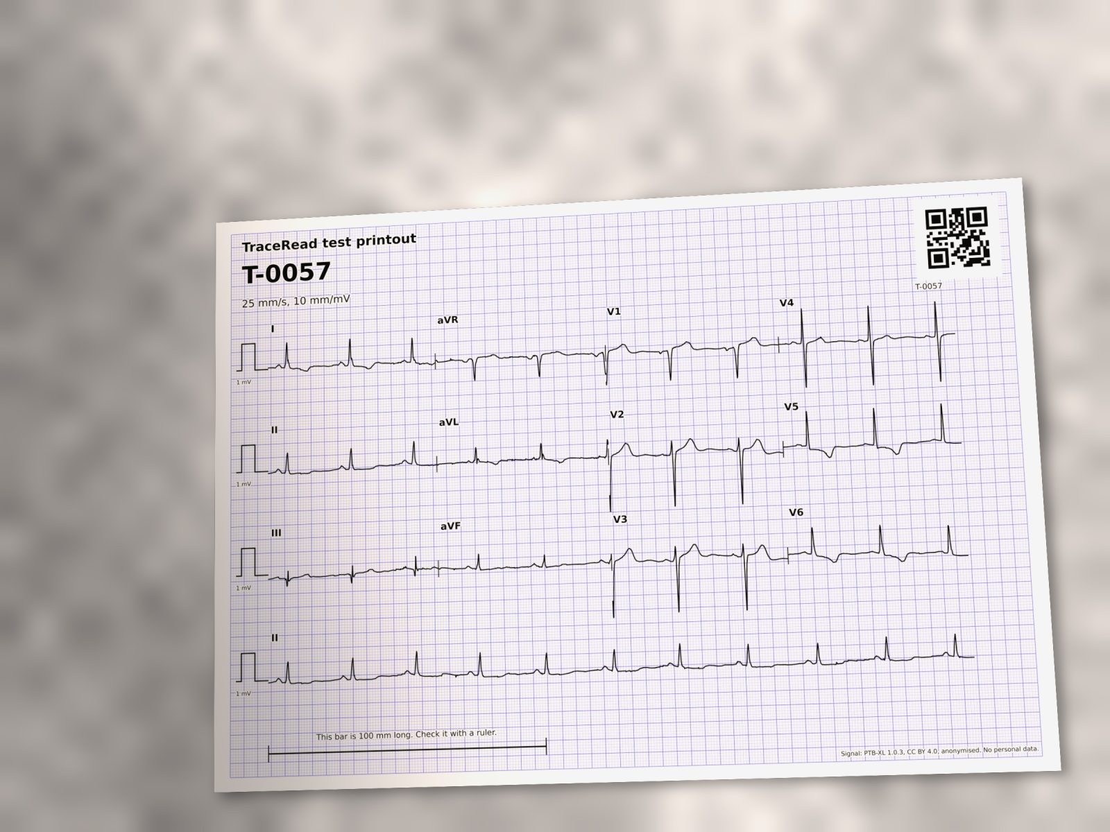
A photo made by computer of a printed public research ECG

Step 1

Take the photo

The technician photographs the printout with a phone. The phone checks sharpness, glare, light and framing first.

What goes in

  • The printed 12 lead ECG
  • A phone with a browser

What comes out

  • One photo near 1.5 MB
  • The header covered on the phone
The page TraceRead AI found, outlined in red

Step 2

Find the page

TraceRead AI finds the four corners of the paper and how it is turned, then fixed code measures the printed grid.

What goes in

  • The photo

What comes out

  • The page corners
  • Paper size and scale
The flattened page

3x4 with one lead II strip, pulses read 4 of 4

Step 3

Read the layout

The calibration pulse at the start of each row tells where each of the 12 leads sits and how tall 1 millivolt is.

What goes in

  • The flat page

What comes out

  • The layout, such as 3x4
  • Pulses read, such as 4 of 4
The flattened page with every rebuilt lead in red

Step 4

Rebuild the trace

TraceRead AI follows the ink of every lead and turns it into a digital signal, 500 points a second, drawn back in red.

What goes in

  • The flat page
  • The layout

What comes out

  • 12 rebuilt leads
  • A match score per lead
I
aVR
V1
V4
II
aVL
V2
V5
III
aVF
V3
V6
II strip

Step 5

Map the seconds

A printout shows most leads for only 2.5 seconds. The coverage map marks exactly which seconds each lead has.

What goes in

  • The rebuilt leads

What comes out

  • A coverage map
  • Nothing missing read as flat
Photo usable

The layout match score was 0.981 (a photo is held back when the score is below 0.888) and 4 of 4 calibration pulses were read. The AI's own uncertainty about the five main heart groups was 0.306 (a photo is held back above 0.497). Neither check fired, so the photo is used.

Step 6

Hold or go

A fixed rule checks the pulses, the layout match and cut leads. If the rebuild looks unreliable, it asks for a new photo.

What goes in

  • The checks above

What comes out

  • Usable, or held for a retake
Heart rate
63 a min
PR interval
136 ms
QRS width
88 ms
QT interval
406 ms
QTc
414 ms
Heart axis
20 deg

Step 7

Measure the beat

Fixed code measures heart rate, PR, QRS, QT, QTc and heart axis on all leads together.

What goes in

  • The rebuilt leads

What comes out

  • Six measures
  • Sinus rhythm flagged

    95
  • Left ventricular hypertrophy flagged

    60
  • Unspecific reduced blood flow pattern flagged

    56
  • Reduced blood flow pattern, front and side flagged

    27
  • ST depression flagged

    19

Step 8

Score the patterns

TraceRead AI scores 71 patterns of rhythm, conduction, thickened muscle and ST and T changes, and flags those past their line.

What goes in

  • The leads and the coverage map

What comes out

  • Patterns with scores

A doctor checks every line.

The AI first read never reaches a patient on its own. Do not rely on it alone in an emergency. A normal AI first read does not rule out heart disease.

Step 9

Your doctor decides

The doctor checks the red line against the paper, every number and every flag, then decides. Nothing reaches a patient before that.

What goes in

  • The AI first read

What comes out

  • A decision by a qualified doctor
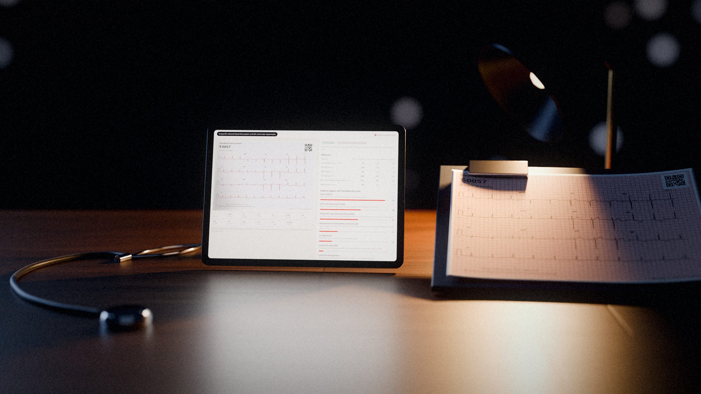
Rendered scene of a dark desk at night, lit mainly by a tablet showing the AI first read of an ECG, with a printed ECG on a clipboard, a stethoscope and a warm desk lamp behind.

Your doctor stays in charge.

At a glance

Numbers you can check.

Every figure says how many ECGs it was measured on and where they came from: public research ECGs from Germany, China and Brazil, and photos of printed ECGs made by TraceRead's photo simulator.

0.908

Trace match

Median match between the rebuilt trace and the original recording, 500 photos made by computer.

20/29

Patterns from China

Patterns scoring 0.90 or more on 45,152 ECGs TraceRead AI never trained on.

7.85 s

Time to read

95 in 100 ECGs read within this on a 2 vCPU server in AWS Cape Town, 97 photos.

16.8 ms

QTc difference

Average difference from a reference ECG program on 2,198 test ECGs.

Safety and privacy

A doctor stays in charge, and patient data stays protected.

An AI first read is not the last word.

  • A qualified doctor reviews every AI first read. Nothing reaches a patient until a doctor signs it.
  • Do not rely on TraceRead alone in an emergency.
  • A normal AI first read does not rule out heart disease.
  • For adults, 18 and over.
Clinical safety

How patient data is handled

  • Patient data stays in the AWS Cape Town region.
  • It is never sent to an outside AI service.
  • TraceRead needs age and sex only, never a name.
  • The printed header is covered on the phone, before the photo leaves it.
  • TraceRead staff cannot see patient data by default.
Privacy and security

Pricing

Pay per AI first read, in naira.

Every plan includes every feature on this site. Your own doctors review and sign.

Pay as you go

₦1,500

per AI first read, about $1.10

  • Every feature on this site
  • Your own doctors review and sign
  • No monthly fee

Centre

₦120,000

a month, about $88

  • 100 AI first reads included
  • Then ₦1,200 per extra read (about $0.88)
  • Every feature on this site
  • Your own doctors review and sign

HMOs and groups

Volume price

for many sites, priced per AI first read

  • One price across all your sites
  • Every feature on this site
  • Your own doctors or medical officers review

Dollar amounts are approximate, at ₦1,370 to the dollar.

Questions

Common questions

What does TraceRead do?

You take a phone photo of a printed 12 lead ECG. TraceRead AI finds the page, rebuilds each of the 12 tracings as a digital signal, measures it and lists the patterns it sees, each with a score from 0 to 100. A doctor then checks that AI first read.

Does TraceRead decide what is wrong with the patient?

No. TraceRead gives an AI first read for a qualified doctor to review. Nothing reaches a patient until a doctor has checked and signed it. A normal AI first read does not rule out heart disease, and TraceRead must not be relied on alone in an emergency.

Which ECGs can it read?

Printed 12 lead ECGs from adults (18 and over), at the usual 25 mm a second and 10 mm per millivolt, laid out as 3x4, 6x2 or 12x1, on A4 or US Letter paper.

Do we need to install anything?

No. The capture app runs in the phone's web browser. It checks each photo before it is sent and keeps photos on the phone until the network is back.

Where do your accuracy numbers come from?

From public research ECGs from Germany (PTB-XL), China (Chapman Shaoxing Ningbo) and Brazil (CODE-15%), and from photos of printed PTB-XL ECGs made by TraceRead's own photo simulator. Each number on this site says how many ECGs it was measured on.

Does TraceRead use a chatbot to write anything?

No. No generative AI writes clinical text. Every pattern name comes from a fixed list, and every measurement is a number from fixed code.

Where is patient data kept?

Patient data stays in the AWS Cape Town region, and it is never sent to an outside AI service. TraceRead needs only the patient's age and sex, never a name. TraceRead staff cannot see patient data by default.

How much does it cost?

₦1,500 per AI first read, or ₦120,000 a month for 100 reads and ₦1,200 for each read after that. HMOs and groups with many sites get one volume price.

See TraceRead read a paper ECG

We show you TraceRead reading public research ECGs, step by step, and talk through how your team prints, photographs and reads ECGs today.